Where is the importance of high-definition imaging for medical precision tungsten contrast catheters in surgery?
1. The principle of high-definition imaging
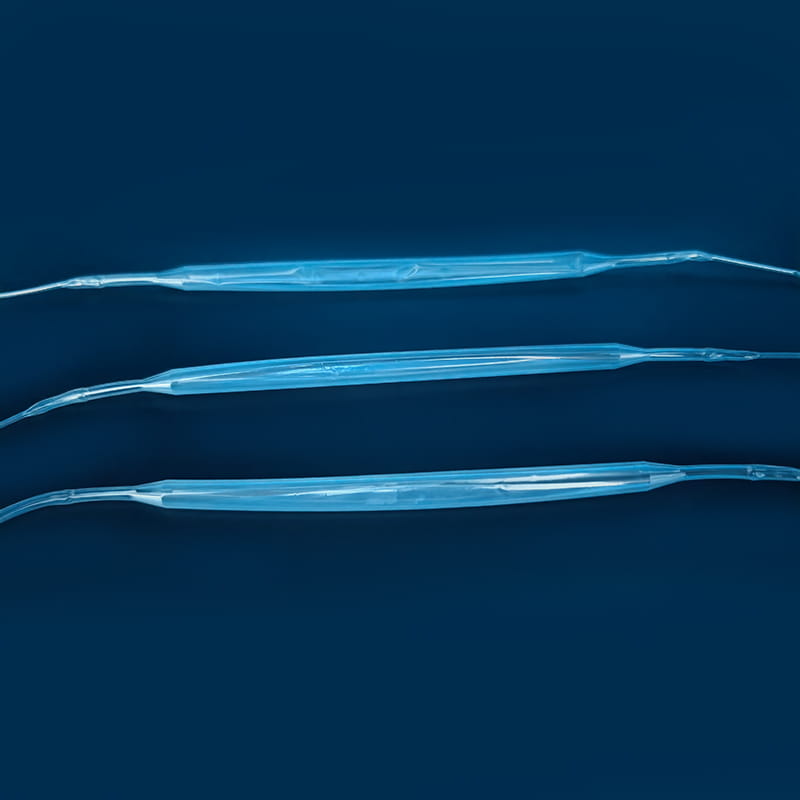
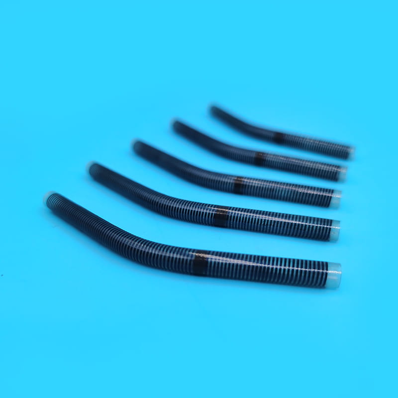
The core of high-definition imaging of medical precision tungsten contrast catheters lies in the unique physical properties of tungsten materials. Tungsten has a high atomic number, which makes it extremely absorbent of X-rays. During angiography or interventional surgery, when X-rays penetrate human tissue and irradiate the contrast catheter containing tungsten markers, the tungsten markers will absorb a large amount of X-ray energy. Compared with surrounding tissues and other developing materials, tungsten markers form a strong signal contrast during X-ray imaging, thus presenting extremely clear images on imaging equipment.
The formation of this clear image is due to the perfect combination of X-ray imaging principles and tungsten material properties. Among the developing materials used in traditional contrast catheters, due to their relatively weak ability to absorb X-rays, the signal contrast is not obvious during imaging, resulting in poor image clarity. The application of tungsten markers has fundamentally changed this situation, providing doctors with clearer and more accurate catheter position information.
II. Navigation tool in complex vascular anatomy
The human vascular system has a complex structure, especially in the cerebral blood vessels, coronary arteries and other parts, where the blood vessels have numerous branches and tortuous directions. In these complex vascular anatomical structures, the high-definition imaging of medical precision tungsten angiography catheters plays a vital navigation role.
In cerebral angiography surgery, the diameter of cerebral blood vessels is slender and the branches are intricate, which requires extremely high accuracy in catheter positioning. A slight deviation may not only fail to obtain accurate diagnostic information, but may also cause damage to fragile cerebral blood vessels. With high-definition imaging, medical precision tungsten angiography catheters allow doctors to clearly see the direction and position of the catheter in X-ray images. Based on the clear images, doctors can accurately judge whether the catheter has entered the target cerebral blood vessel branch, adjust the direction and depth of the catheter in time, and guide the catheter to the lesion site smoothly. This precise navigation capability greatly improves the accuracy and success rate of cerebral angiography, and provides a reliable basis for the diagnosis of cerebrovascular diseases.
In coronary angiography, the beating of the heart and the special morphology of the coronary arteries increase the difficulty of catheter operation. The high-definition imaging of medical precision tungsten angiography catheters enables doctors to clearly observe the position and status of the catheter in the coronary artery during dynamic heart movement. Doctors can accurately determine whether the catheter has successfully passed through the stenosis of the coronary artery, and provide accurate position references for subsequent possible interventional treatments, such as stent implantation, to ensure the smooth implementation of the treatment plan.
3. Key guarantees for reducing surgical risks
During the operation, misjudgment of the catheter position may lead to a series of serious consequences, such as vascular perforation, rupture, and damage to surrounding important tissues and organs. The high-definition imaging of medical precision tungsten angiography catheters provides doctors with detailed and accurate catheter position information, effectively avoiding surgical risks caused by misjudgment of the catheter position.
In interventional treatment surgery, doctors need to accurately deliver the catheter to the lesion site and deliver therapeutic devices or drugs through the catheter. If the catheter position cannot be clearly determined, the therapeutic device or drug may be delivered to the wrong location, affecting the treatment effect, or even causing harm to the patient. The high-definition imaging tungsten angiography catheter allows doctors to accurately grasp the catheter position in real time during the operation, ensuring that the therapeutic device or drug is accurately delivered to the lesion. In tumor embolization therapy, doctors can accurately inject embolic materials into the blood vessels supplying the tumor based on clear catheter images, effectively blocking the blood supply of the tumor, while preventing the embolic materials from flowing into normal blood vessels by mistake, reducing damage to normal tissues.
In some complex operations that require repeated adjustment of the catheter position, high-definition imaging can help doctors operate the catheter more accurately and reduce damage to the blood vessels caused by repeated adjustments of the catheter. Clear images allow doctors to clearly understand the contact between the catheter and the blood vessel wall, adjust the operation force and direction in time, reduce the risk of complications such as vascular intimal damage and dissection formation, and ensure the safety and smooth progress of the operation.
Fourth, the core elements to improve the success rate of surgery
In medical practice, the success rate of surgery is directly related to the patient's rehabilitation effect and life health. The high-definition imaging of medical precision tungsten angiography catheters provides strong support for the success of the operation and becomes the core element to improve the success rate of the operation.
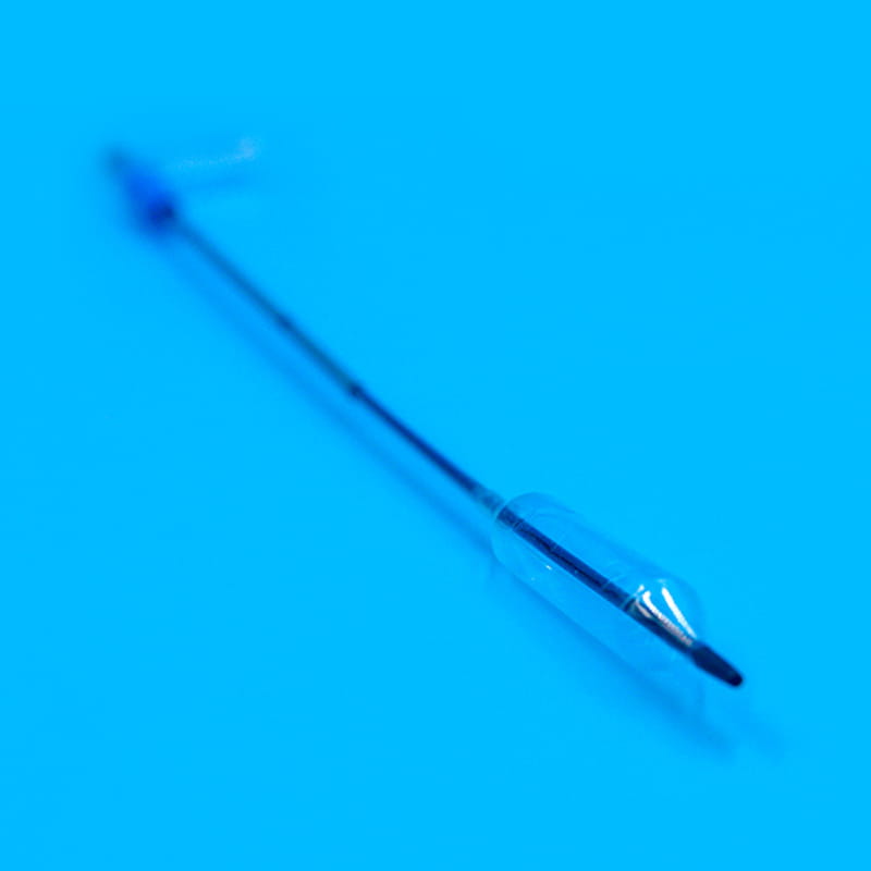
In interventional treatments such as angioplasty, accurate catheter positioning is the key prerequisite for the success of the operation. High-definition imaging enables doctors to more accurately guide balloon catheters or stent delivery systems to the narrowed parts of the blood vessels. Doctors can accurately judge the position and expansion degree of the stent release based on clear images, ensure that the stent fits closely with the blood vessel wall, effectively expand the narrowed blood vessels, and restore blood vessel patency. Compared with traditional angiography catheters, the use of high-definition imaging tungsten angiography catheters for angioplasty can significantly improve the accuracy and success rate of the operation and reduce the incidence of complications such as postoperative restenosis.
In the treatment of some complex vascular diseases, such as multi-vessel lesions and severe vascular tortuosity, the advantages of high-definition imaging are more prominent. Doctors can use clear catheter images to develop more reasonable surgical plans and choose more appropriate treatment equipment and operation methods. When facing multi-branch coronary artery lesions, doctors can accurately evaluate the conditions of each diseased blood vessel based on clear angiography images, reasonably arrange the order and number of stent implants, improve the overall success rate of the operation, and improve the patient's prognosis.
For more information, please call us at +86-18913710126 or email us at [email protected].
Introduction In medical practice, particularly in post-surgical care, body cavity effusion, orthoped...
In the medical field, the safety, stability, and efficiency of fluid transmission are directly relat...
Introduction In modern medical procedures, particularly those involving minimally invasive surgery o...
Vascular interventional procedures are integral to modern cardiovascular medicine, particularly when...
Introduction Single-lumen endobronchial tubes are a critical component of respiratory therapy, espec...
The medical industry is increasingly relying on advanced materials for various applications, and one...


